Once you begin looking closely at an iris, you realize very quickly that it is not simply a flat circle of color. There are fibers, openings, darker and lighter areas, pigment spots, rings, folds, and patterns that can look completely different from one person to another.
This is part of what makes iridology so fascinating to study. It is also where things can become confusing, because many of the same visible features have both an anatomical description and a traditional iridology interpretation. Those two things are not automatically the same.
In holistic iridology, practitioners may use iris features as part of a conversation about constitution, tendencies, and overall wellness patterns. Modern ophthalmology, however, does
not support using those markings to diagnose disease elsewhere in the body. Current reviews continue to find that iridology has not demonstrated reliable diagnostic accuracy as a medical screening method. PubMed
That distinction is going to come up a lot in this article, because I think we can appreciate how interesting the iris is without asking it to tell us something it has not been scientifically shown to tell us.
The Iris Really Does Have Patterns
Many of the markings discussed in iridology are not imaginary. They are genuine structures that can be seen on the surface of the iris.
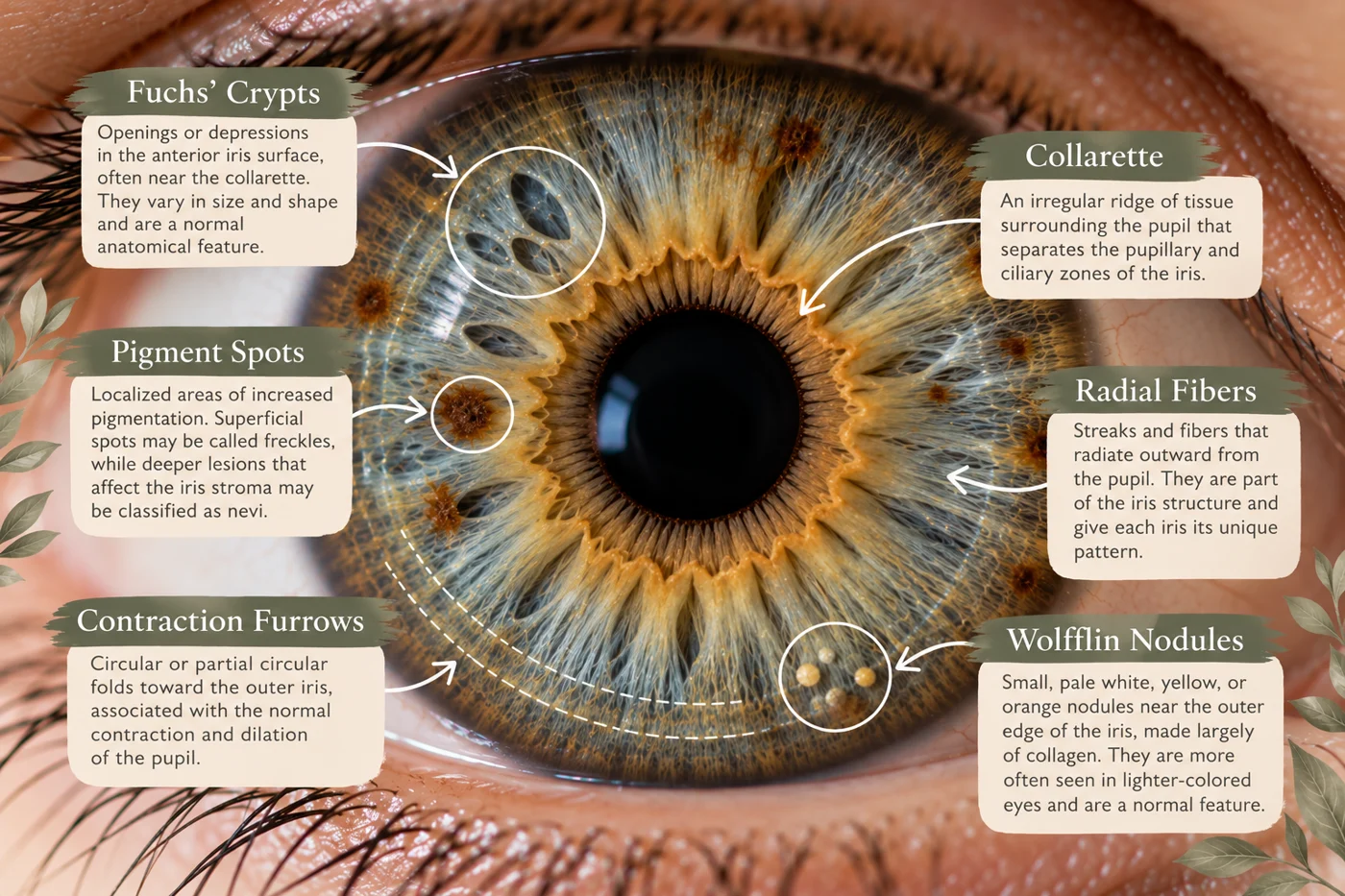
Researchers describe features including Fuchs' crypts, contraction furrows, pigment spots, Wolfflin nodules, radial streaks, and the collarette. These features vary considerably between individuals, and studies have found differences in their frequency among populations with different ancestry and iris characteristics. PubMed Central (PMC)
The important question is not whether the markings exist.
They do.
The question is what conclusions we can reasonably draw from them.
Those Circular Rings: Contraction Furrows
One of the easiest iris features to notice is a series of circular or partial circular grooves toward the outer portion of the iris. In anatomical literature, these are known as contraction furrows.
They form as folds within the iris and are associated with the repeated contraction and dilation of the pupil. They may appear as complete-looking rings or as shorter, staggered arcs around portions of the iris. Researchers also believe the thickness and density of iris tissue influence how visible they become. PubMed Central (PMC)
Within some traditional iridology systems, contraction furrows have historically been called “nerve rings” or “stress rings.” Practitioners may interpret their number, depth, location, or pattern as part of a constitutional discussion about nervous-system tension or how someone responds to stress.
That is an iridology interpretation, however—not evidence that someone has chronic stress because rings appear in their iris. Anatomically, contraction furrows are common physical folds of the iris, and current evidence does not establish them as a diagnostic measure of a person's stress level or nervous-system health. PubMed Central (PMC)
That does not prevent an iridologist from asking about stress during a holistic assessment. It simply means the conversation should not become, “You have three rings, therefore your nervous system is unhealthy.”
Crypts and Lacunae: The Little Open Spaces
Another feature people often notice is an area where the normal iris texture appears more open. These openings can vary greatly in size and shape.
In anatomy, Fuchs' crypts are openings in the anterior portion of the iris that develop during formation of the eye. They often appear around the collarette and can look somewhat like small windows or diamond-shaped spaces within the iris fibers. They are among the normal structural variations researchers study when examining iris patterns. PubMed Central (PMC)
Iridology uses additional terminology such as lacunae, and different schools may classify these openings based on shape, depth, position, whether they appear open or closed, and how they relate to surrounding iris structures. Traditional systems may interpret them as part of a person's constitutional tissue pattern.
This is where I think language matters enormously. An iridologist can say, “Within this iridology system, this type of opening has traditionally been interpreted as a constitutional characteristic.” That is very different from telling someone that an opening proves an organ is weak or diseased.
The visible structure is real.
The medical diagnosis is not something we can derive from it.
Pigment Spots: Color Within Color
Pigment spots are another feature that immediately catches the eye, especially in a lighter iris. From an anatomical perspective, pigment spots are localized areas of increased pigmentation.
Researchers distinguish superficial iris freckles from deeper nevi that can affect the underlying iris stroma. Iris pigmentation and pattern are influenced substantially by genetics, and freckles can also be associated with environmental factors such as sun exposure. PubMed Central (PMC)
Traditional iridology systems often devote considerable attention to pigment. Practitioners may look at the shade, location, distribution, or whether pigment appears concentrated around particular portions of the iris. Various iridology traditions have developed interpretations around those patterns.
Again, I would keep those descriptions firmly in the category of traditional interpretation rather than medical finding.
A brown spot in a particular portion of an iris does not scientifically establish that the corresponding organ on an iridology chart has a problem. Modern reviews specifically identify pigmentation patterns among the features that iridology has attempted to use diagnostically, but the evidence has not supported reliable diagnosis of systemic disease from those patterns. PubMed
There is also an important medical distinction here. A new or changing pigmented lesion in the eye deserves evaluation by an eye-care professional. Ophthalmology recognizes a range of benign and, less commonly, concerning iris lesions, and those are evaluated using medical eye examination rather than an iridology map. PubMed
The Collarette: That Uneven Ring Around the Pupil
If you look closely at many irises, you can see an irregular circular structure surrounding the pupil. This is the collarette, and unlike some wellness terminology, that is an actual anatomical landmark.
The collarette separates the more central pupillary portion of the iris from the outer ciliary portion. It develops during formation of the eye and is commonly used as a reference point when describing other iris structures. PubMed Central (PMC)
In holistic iridology, practitioners may study the shape, width, regularity, prominence, and openings around the collarette as part of their constitutional interpretation. Some systems connect the area with digestive or autonomic patterns.
Those associations belong to iridology teaching systems. The physical collarette itself is real anatomy, but its shape has not been validated as a way to diagnose digestive disorders or determine how well someone's gastrointestinal system is functioning. PubMed
This is a good example of why I think holistic iridology is most responsibly used as an observational framework, not a medical test.
Radial Lines and Fiber Patterns
The iris also contains radial structures extending outward from the pupil. Some of what we see reflects the organization of the underlying stromal tissue and blood vessels. Researchers describe radial streaks and a loose mesh of collagen fibers, pigment-producing cells, and other structures that contribute to the unique appearance of each person's iris. PubMed Central (PMC)
Iridology systems often pay attention to the overall appearance of these fibers: how tightly arranged they seem, whether there are areas of separation, and whether particular lines appear to interrupt the otherwise radial pattern.
Traditional practitioners may use terms such as radial furrows, transversals, rarefactions, or fiber density, depending on the school of iridology being taught. These observations may then contribute to a broader constitutional interpretation.
This is another area where I would resist turning a visual feature into a declaration about someone's health. The iris contains enormous natural variation, and some of the patterns seen in perfectly healthy eyes are simply part of the structure that makes one iris different from another.
And What About the Little Light-Colored Dots?
Some lighter irises contain small pale or whitish spots around the outer portion of the iris. One anatomical term you may encounter is Wolfflin nodules.
These are small accumulations of collagen that typically occur toward the outer iris and are more visible in lighter-colored eyes. They are another recognized iris feature found in healthy people. PubMed Central (PMC)
Various iridology traditions have also interpreted light-colored spots or clusters according to their own systems. Older iridology literature may use terminology that sounds much more medically certain than I would be comfortable using.
This is exactly why I think learning the anatomy alongside iridology is so valuable. The more we understand what we are actually looking at, the easier it becomes to separate observation from assumption.
One Mark Should Never Tell the Whole Story
Even within holistic iridology, I do not think one mark should become the entire assessment.
Two people can have similar-looking iris structures and completely different lifestyles, histories, symptoms, stress levels, diets, sleep patterns, environments, and health experiences. Looking at one crypt, one pigment spot, or one contraction furrow in isolation strips away all of that context.
A more thoughtful approach is to observe the overall iris pattern and then use it as one piece of a much larger conversation.
What has the person already noticed?
How do they eat?
How are they sleeping?
What does stress look like?
How is digestion?
What does movement look like?
What has changed recently?
And when there is a medical concern, what have appropriate medical evaluation and testing actually shown?
The iris should not override the rest of the person.
Why Different Iridologists May Use Different Terms
Something else readers may notice when researching iridology is that terminology is not completely standardized.
There are classical American systems, European traditions, constitutional iridology, holistic and multidimensional approaches, and other schools that may describe or interpret similar features differently. Books and training programs may use different names for related markings or assign different significance to them.
Even dedicated iridology texts separate topics such as contraction furrows, crypts, pigmentation, lacunae, collarette characteristics, pupillary changes, and constitutional types into distinct interpretive systems. Iridology Clinic
That variability is another reason I do not think we should present an iridology interpretation as though it were an objective laboratory result.
It is a framework being applied to an observation.
That distinction protects the person receiving the information and keeps the practitioner honest about what the practice can and cannot establish.
Can Iris Markings Change?
The iris is often spoken about as though it is a constantly changing health report, but that needs nuance too.
Much of an individual's iris structure is strongly influenced by genetics and development. Features such as crypts, furrows, pigmentation patterns, and overall architecture contribute to the iris's highly individual appearance. Research has demonstrated meaningful genetic influences on both iris color and pattern. PubMed
Some visible eye findings can change over time, including certain pigmentary features or actual eye disease, but that does not mean an iris should be expected to redraw itself every time someone's wellness changes.
If a spot or lesion appears new or visibly changes, especially if it is accompanied by pain, redness, vision changes, pupil changes, or other eye symptoms, that belongs with an optometrist or ophthalmologist rather than being interpreted as a wellness “detox sign.” PubMed
What I Want Someone to Take Away From an Iris Assessment
I do not want someone leaving an assessment afraid of their eyes.
I do not want them staring at a dark spot wondering whether an organ is failing or counting rings and deciding they are too stressed to function.
I would rather they come away with curiosity.
Maybe an iris characteristic becomes the starting point for a conversation about stress,
hydration, digestion, movement, nourishment, or rest. Maybe that conversation confirms something they had already been noticing. Maybe it simply makes them pay closer attention to one of their everyday foundations.
And sometimes the appropriate conclusion is simply:
That is part of what makes your iris uniquely yours.
That is allowed too.
Observe
The next time you look closely at your eyes, notice how much structure is actually there. Look at the fibers. Notice the collarette around the pupil. Look for variations in pigment, little openings in the texture, and the circular folds that may appear around the outside.
You do not have to assign a problem to every feature you see.
The iris is an incredibly detailed piece of human anatomy, and simply learning to observe it can be fascinating.
Within holistic iridology, those patterns can become part of a larger conversation about constitution and wellness. But they should never become a substitute for medical testing, an eye examination, or the story the person sitting in front of us is actually telling.
The marking is an observation. What we do with that observation is where responsibility begins.
Curious About Your Own Iris?
As I continue my education in holistic iridology, I’m offering complimentary student practice readings. These educational readings are an opportunity to explore visible iris patterns and learn how they’re traditionally interpreted in holistic iridology.
Explore Free Holistic Iris ReadingsFor education and student practice only. Iridology is not a medically validated diagnostic method and does not replace medical care or an eye examination.
A Note About Holistic Iridology
Holistic iridology is a non-diagnostic wellness assessment practice. Traditional iridology interpretations of iris markings have not been scientifically validated as a method for diagnosing or screening for disease. New or changing iris spots, eye pain, redness, pupil changes, vision changes, or other concerning eye findings should be evaluated by an appropriately qualified eye-care professional.
References
Iuliano L, Veritti D, Lanzetta P. Iridology: Biological plausibility, clinical evidence, and implications for ophthalmic practice. 2026. This recent ophthalmology review concluded that current evidence does not support iridology as a diagnostic or screening method. PubMed
Ernst E. Iridology: A systematic review. 1999. A review of blinded studies evaluating iridology's diagnostic validity found that the evidence did not support it as a diagnostic method. PubMed
Edwards M, et al. Analysis of iris surface features in populations of diverse ancestry. Describes normal iris structures including Fuchs' crypts, contraction furrows, pigment spots, Wolfflin nodules, and the collarette. PubMed Central (PMC)
Sturm RA, Larsson M. Genetics of human iris colour and patterns. Reviews the genetics and biological basis of human iris color and pattern variation. PubMed
Daneshvar K, et al. Iris lesions: A review. 2026. Reviews the range of iris lesions recognized in ophthalmology and reinforces the importance of medical evaluation of concerning or changing lesions